
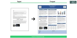
- Services
- Publication Support Services
-

Publication Support Service
-
- Editing & Translation
-
Editing and Translation Service
-
- Research Services
-
Research Services
-
- Physician Writing
-
Physician Writing Service
-
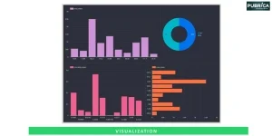
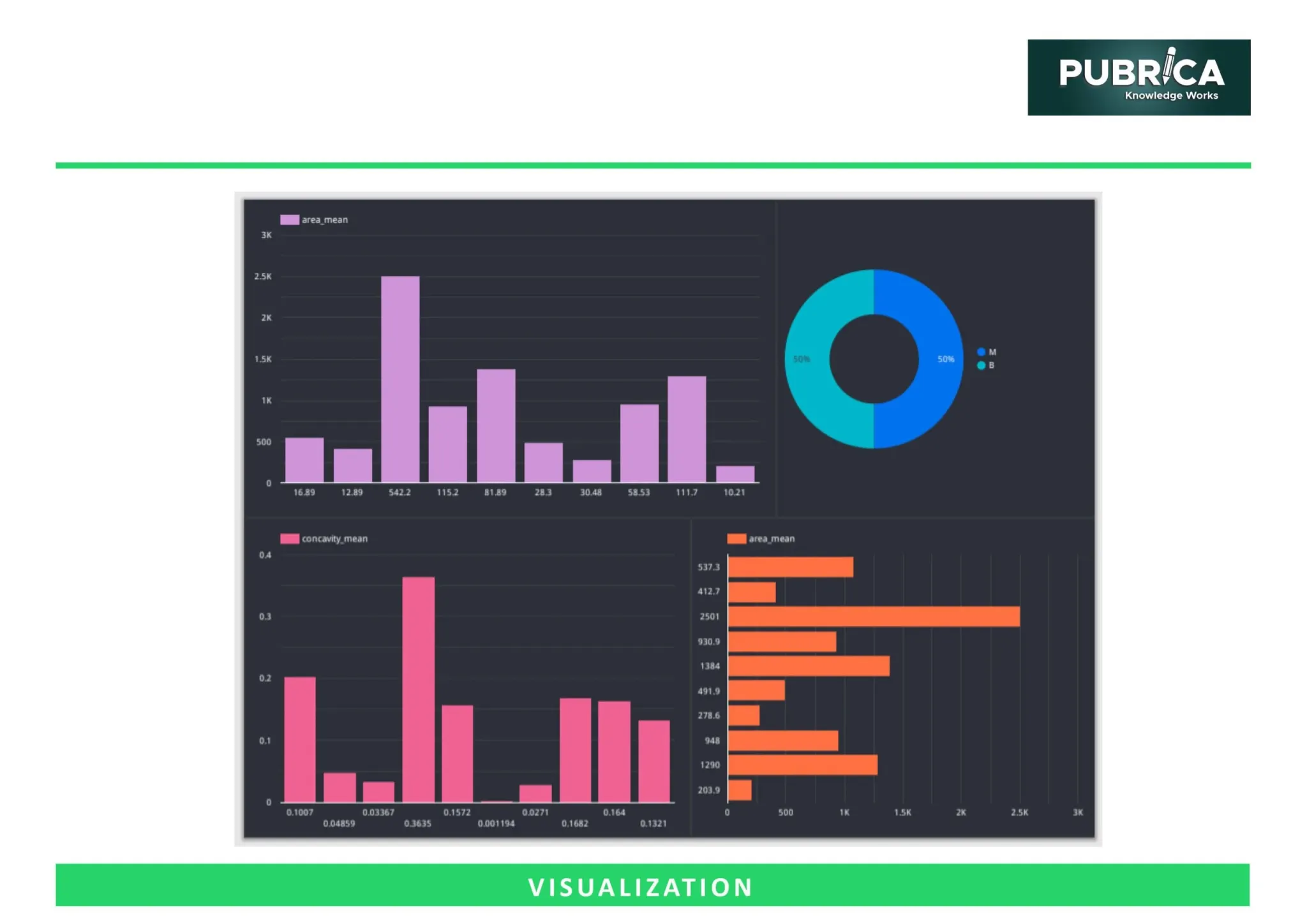
- Statistical Analyses
-
Statistical Analyses
-
- AI and ML Services
-
AI and ML Services
-
- Medical Writing
-
Medical Writing
-
- Research Impact
-
Research Impact
-
- Medical & Scientific Communication
-

Medical & Scientific Communication
-
- Academic Editorial Services
-
Academic Editorial Services
-
- Educational Editorial Service
-
Education Editorial Services
-
- Publication Support Services

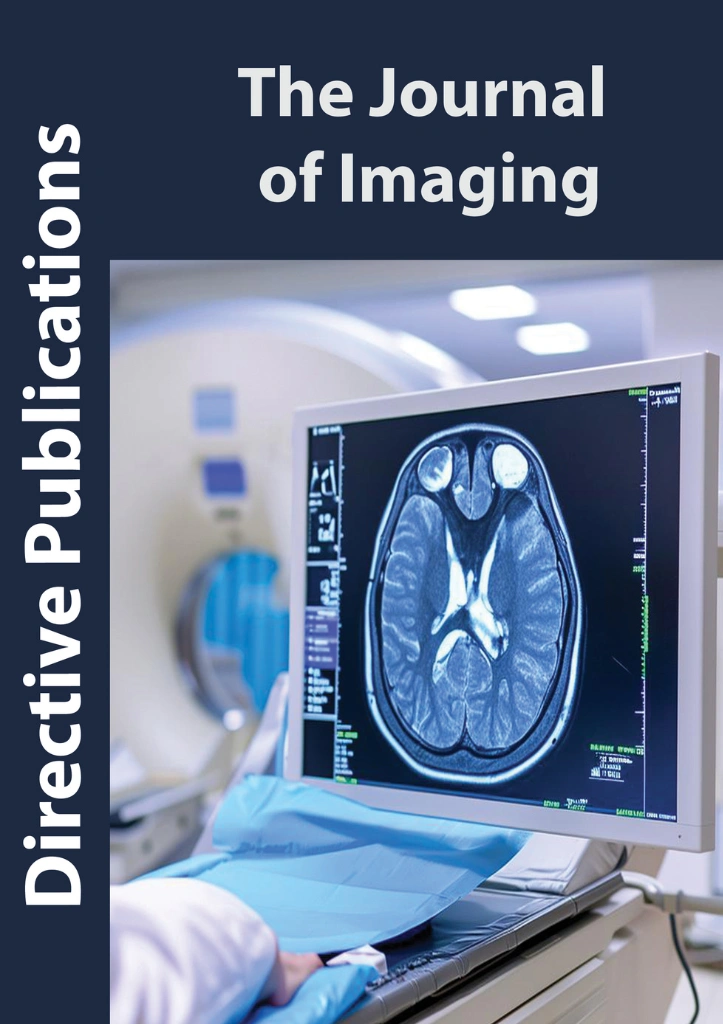
X-ray Imaging
It is one of the oldest and most widely used diagnostic tools. It utilizes ionizing radiation to produce images of bones and certain tissues. Innovations such as digital radiography and computed tomography (CT) have significantly improved image quality, reduced exposure, and expanded clinical applications.

Computed Tomography
(CT)
CT combines multiple X-ray images taken from different angles to produce cross-sectional views of the body. This technique allows precise visualization of internal organs, blood vessels, and skeletal structures. Modern CT imaging is critical for trauma assessment, cancer detection, and cardiovascular evaluations.

Magnetic Resonance Imaging (MRI)
MRI employs powerful magnetic fields and radio waves to generate detailed images of soft tissues. Unlike X-rays, MRI does not involve ionizing radiation, making it safe for repeated use. MRI is particularly valuable in neurological studies, musculoskeletal assessments, and cardiac imaging.

Ultrasound Imaging
Ultrasound uses high-frequency sound waves to create real-time images of organs and tissues. It is non-invasive, portable, and widely used in obstetrics, cardiology, and abdominal diagnostics. Advanced techniques like Doppler ultrasound can visualize blood flow and vascular health.

Nuclear Medicine and PET Imaging
Nuclear imaging involves the use of radioactive tracers to evaluate physiological processes. Positron Emission Tomography (PET) and Single Photon Emission Computed Tomography (SPECT) provide functional insights into metabolism, organ function, and disease activity, complementing structural imaging methods.

Optical Imaging
Optical imaging techniques, including fluorescence and bioluminescence imaging, are predominantly used in research and preclinical studies. They enable the visualization of cellular and molecular processes in real-time, advancing our understanding of disease mechanisms.


The editorial support we received was exceptional. The team improved the clarity of our MRI-based diagnostic study without changing the scientific meaning. Their expertise in image reconstruction and data interpretation greatly strengthened our manuscript, and it was accepted in a high-impact journal on the first submission.


The editors demonstrated excellent knowledge of radiomics and machine learning in medical imaging. Their precise edits improved the flow, scientific tone, and alignment with journal guidelines. Thanks to their guidance, our manuscript was published within two review cycles.


I was impressed with their deep understanding of PET-CT analysis and quantitative imaging. They not only enhanced the language but also identified missing methodological details that helped us significantly improve our paper before peer review. Highly recommended for biomedical imaging publications.


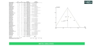
"Pubrica’s team provided exceptional support throughout my meta-analysis cardiovascular drug efficacy. Their adherence to PRISMA guidelines and attention to statistical detail helped me publish in the European Heart Journal. Highly recommended"
— Dr. Anna Müller
Cardiologist, University Hospital Munich, Germany

"The meta-analysis manuscript I co-authored with Pubrica’s experts was accepted by BMC Public Health without major revisions. Their data synthesis and transparent methodology were critical to this success."
— Dr. Rohan Mehta
Public Health Researcher,
All India Institute of Medical Sciences (AIIMS), India

"Thanks to Pubrica’s guidance, our meta-analysis on paediatric nutrition was published in The Lancet Child & Adolescent Health. The methodological rigor and rewriting support were key contributors to the paper’s clarity and impact."
— Dr. Luis Fernández
Pediatrician & Research Fellow, University of Barcelona, Spain


How to Structure Case Reports and Review Articles for Medical Journals
Medical journals expect a structure for case reports and review articles, with clear objectives....

How Should Physicians Choose the Right Journal for Submitting a Case...
Publishing a case report involves more than clinical knowledge; it also demands strategic journal ....

How Physicians Can Write Clear and Impactful Patient Education Materials
Effective patient education materials (PEMs) are crucial for promoting health literacy, enhancing....